
Bluesky: Diffraction images from flow cytometer

diffraction flow cytometer

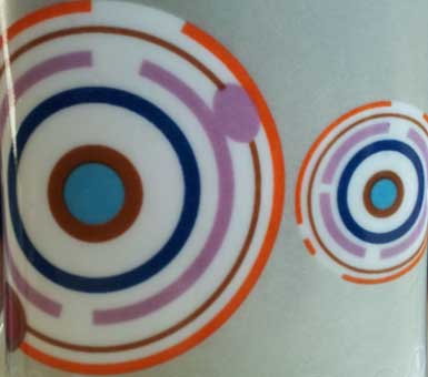
Flow cytometry typically flows particles or cells through a laminar flow cell into an interrogating laser beam to be read out as flourescent signal with a photomultiplier or photodiode. The read out is typically the integrated intensity or a two dimensional projection of a cell. The residence time is in order of 10 micro-seconds. Cellular high resolution microscopy in contrast acquires 2D or 3D image of the cell but the process is excruciatingly slow. A researcher at East Carolina university, Dr Xin-Hua Hu has been analyzing diffraction images of cells passing through flow cytometer. The concept being that you will be able to acquire multi-dimensional images very quickly that will have information about the shape, location and other information about the cellular components. The diffraction images do not look like any images that you have seen before of cells and instead look like clouds in the sky. However the anticipation is that these images are unique enough to distinguish and quantitate one cell from the other.

http://bmlaser.physics.ecu.edu/

Similar Posts

  • Imaging through rain.

    Start with the application: A set of headlights that can enable the driver to see through the falling rain. Typically, it is difficult to see through rain because water droplets reflect the light that is reflected back from the water droplet. However, rain is not constant and it should be possible to time the light…

  • Image analysis – outsourced, simple and free

     Image analysis used to be very labor-intensive operation. It required programmers to understand what was being analyzed and then developing algorithms to specifically find the areas of interest. Then came Machine learning and Artificial intelligence tools. These found and analyzed the data but now you needed different set of programmers and they needed more sample…

  • 3D microscopy

    Super resolution microscopy One of the problems with Quantum dots and some fluorescent proteins/molecules is that they blink. And they all photo-bleach. Bayesian analysis of blinking and bleaching or 3B microscopy analyzes images using Hidden Markov Model (HMM) allows useful information to be extracted from the data. Dr Susan Cox has come up with unique…

  • Bioluminiscent imaging

    Bio-luminescent imaging Hair growth has been on ongoing research topic It is thought that stem cells can regenerate hair follicles when injected into animals and humans. However, this process has not been imaged very clearly. A group of scientists from Kyungpook National University School of Medicine, Daegu, Republic of Korea have used bioluminescent imaging to…