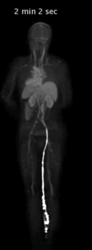
Human PET (Positron Emission Tomography) scanning is used frequently to determine the metabolism of glucose in the body which help find cancer and also for finding specific molecules using specific PET probes.
However, PET scanners are expensive to operate, expose the body to radiation and also require long amounts of time for the patient in the scanner while images are acquired.
Imagine creating a picture of human body that could display PET and CT together and do the scanning all at once. The amazing thing about the scanner is that it enables the PET scan to occur in about 1 minute and also do it with 40x less radiation.
University of California Davis have created just the scanner.
See it on their website at https://explorer.ucdavis.edu/
The video above shows the amazing technology behind the scanner that can capture the full body image in just about a minute. What this will enable is studies that can track the movement of the probe of tracer all through the body.
Wonder if more probes will be developed to complement the scanner?