EXPLORER PET scanner – fast!

whole body image

Human PET (Positron Emission Tomography) scanning is used frequently to determine the metabolism of glucose in the body which help find cancer and also for finding specific molecules using specific PET probes.

However, PET scanners are expensive to operate, expose the body to radiation and also require long amounts of time for the patient in the scanner while images are acquired.

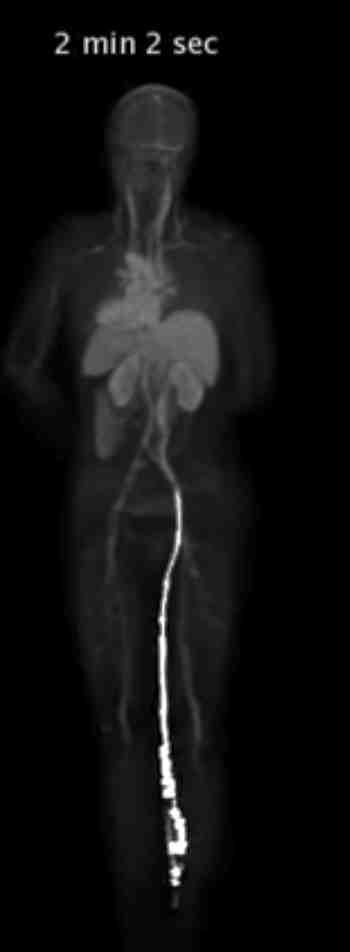
Imagine creating a picture of human body that could display PET and CT together and do the scanning all at once. The amazing thing about the scanner is that it enables the PET scan to occur in about 1 minute and also do it with 40x less radiation.

University of California Davis have created just the scanner.

See it on their website at https://explorer.ucdavis.edu/

The video above shows the amazing technology behind the scanner that can capture the full body image in just about a minute. What this will enable is studies that can track the movement of the probe of tracer all through the body.

Wonder if more probes will be developed to complement the scanner?

Similar Posts

  • Imaging through rain.

    Start with the application: A set of headlights that can enable the driver to see through the falling rain. Typically, it is difficult to see through rain because water droplets reflect the light that is reflected back from the water droplet. However, rain is not constant and it should be possible to time the light…

  • Photoacoustic imaging

    It is difficult to image at high resolution using conventional optics to any reasonable depth below the skin. There are many ways to counteract this and one unique method is called photoacoustic imaging. This method was invented by Lihong Wang at Washington University in St Louis. The phenomenon of acoustic imaging is fascinating use of…

  • Single molecule super resolution microscopy made simple

    Bayes methods have come to microscopy. Researchers have developed the Bayesian analysis of photo-blinking and photo-bleaching in microscopy to deduce the structure that is not usually visible. (Bayesian localization microscopy reveals nanoscale podosome dynamics, Cox, S et al, Nat. Methods 9, 195 (2012)) http://www.nature.com/nmeth/journal/vaop/ncurrent/full/nmeth.1812.html There have been many methods that are used for super resolution…

  • Malaria games

    We think that image processing problems are difficult and interesting but there are others that elude us all the time. Malaria, a mosquito borne illness affects nearly 600,000 patients per day. The diagnosis unfortunately in many countries still uses the traditional Giemsa stain of blood. This requires a pathologist to study the slides and then…