Atomic Force microscopy for eyes

Atomic force microscopy for eyes

Label free Atomic Force Microscope screening

Diabetic retinopathy and scar tissue form on the eye in diabetics causing blindness or visual impairment. This is handled different ways by the doctors. They either use a laser to ablate the vascularization and the scar tissue or go inside the eye and peel off the inner limiting membrane and scar tissue. However, it is difficult to differentiate the retinal cells from the outer membrane. The only technology to separate is to use a dye – but there are no dyes approved in the US.

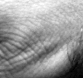
Atomic Force microscopy (AFM) modified with bimodal dual AC mode microscopy has been used by Ivanisevic lab at the North Carolina State to determine the surface structure of the retina. Thus this label free microscopy technique is very useful for determining the structure of the cells on the surface based on their structure and in the absence of any fluorescent or other labeling modalities. This will be used for testing of new dyes that could be used for determining retinal layer from the other layers.

Though AFM is slow and tedious, the resolution is very high and the supplemental information about the surface forces will be important to develop new molecules for identification of retina

https://news.ncsu.edu/2012/06/wms-ivanisevic-eye/

Similar Posts

  • Lens free microscopy

    Lens free microscopy Microscopy whether electron microscopy or with visible light requires lenses. For electron microscopy, lenses incur significant design challenges and add aberrations. A startup in UK uses technology invented by Professor John Rodenberg to remove lenses altogether and compute the image from the diffraction pattern of the sample. This is similar to a…

  • Photoacoustic imaging

    It is difficult to image at high resolution using conventional optics to any reasonable depth below the skin. There are many ways to counteract this and one unique method is called photoacoustic imaging. This method was invented by Lihong Wang at Washington University in St Louis. The phenomenon of acoustic imaging is fascinating use of…

  • Image based Phenotypic screening

    When screening for phenotypes there are many methods that are used. Usin’Life uses artificial intelligence methods to analyze the image data sets but there have other laboratories that use other interesting methods. Dr Lu’s lab published a paper in Nature methods (Nat. Methods 9, 977–980, 2012) that uses microfluidic technology to determine the phenotypic characteristics…

  • Elastomer for surface imaging

    Surface geometry made easy It is extremely difficult to measure the cell surface topology using microscopes or camera’s. Typically, the choice is to use reflected light, refracted light or some unique modification that requires light to enter at correct angles, be altered and then measured with the camera. Typically, setup like that cost hundreds to…

  • Computational Photography in Science

    In the field of imaging there is another branch of image acquisition/analysis/study that is called Computational Photography as distinguished from traditional photography. In traditional photography, with a regular camera, you take a picture of a 3D scene through a camera. This light from the scene is captured as a 2-dimensional representation onto a photo-sensitive surface,…